. DOI: 10.1364/OPTICA.518559")
Sensing deflected wavefronts in fluorescence microscopy. Credit: Optics (2024). DOI: 10.1364/OPTICA.518559
A team led by researchers at HHMI’s Janelia Research Campus has adapted a class of techniques used in astronomy to whiten images of distant galaxies for use in the life sciences, offering biologists a faster and cheaper way to get clearer and sharper microscopic images. The findings are published in the journal Optics.
Astronomers long ago figured out how to make the images their telescopes capture of distant galaxies clearer and sharper. Using techniques that measure how light is distorted by the atmosphere, they can apply corrections to cancel out the aberrations.
Microscopists have adapted these methods to generate sharper images of thick biological samples, which also bend light and create distortions. But these techniques — a class of methods called adaptive optics — are complex, expensive and slow, putting them out of reach for many labs.
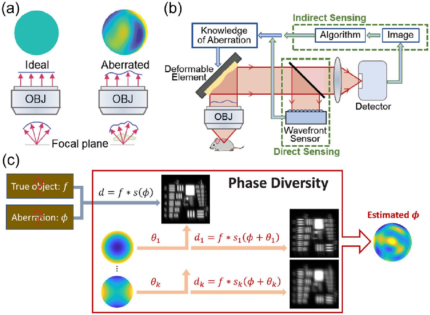
Now, in hopes of making adaptive optics more widely available to biologists, a team led by researchers at HHMI’s Janelia Research Campus has turned their attention to a class of techniques called phase diversity that has been widely used in astronomy, but is new in the life sciences.
These phase diversity methods add additional images with known deviations to a fuzzy image with an unknown deviation, providing enough additional information to decode the original image. Unlike many other adaptive optics techniques, phase diversity does not require any major changes to an imaging system, making it a potentially attractive avenue for microscopy.
To implement the new method, the team first adapted the astronomy algorithm for use in microscopes and validated it with simulations. Next, they built a microscope with a deformable mirror, whose reflective surface can be changed, and two additional lenses—small modifications to an existing microscope that create the familiar aberration. They also improved the software used to perform phase diversity correction.
As a test of their new method, the team showed that they could calibrate the microscope’s deformable mirror 100 times faster than with competing methods. They then showed that the new method could sense and correct for randomly generated aberrations, providing clearer images of fluorescent beads and fixed cells.
The next step is to test the method on real-world samples, including living cells and tissues, and extend its use to more complex microscopes. The team also hopes to make the method more automated and easier to use. They hope the new method, which is faster and cheaper to implement than current techniques, could one day make adaptive optics accessible to more labs, helping biologists see more clearly when they look deep. within the tissues.
More information:
Courtney Johnson et al, Phase diversity-based wavefront sensing for fluorescence microscopy, Optics (2024). DOI: 10.1364/OPTICA.518559
Provided by Howard Hughes Medical Institute
citation: Scientists adapt astronomy method to unblur microscopy images (2024, June 12) retrieved June 13, 2024 from https://phys.org/news/2024-06-scientists-astronomy-method-unblur-microscopy.html
This document is subject to copyright. Except for any fair agreement for study or private research purposes, no part may be reproduced without written permission. The content is provided for informational purposes only.